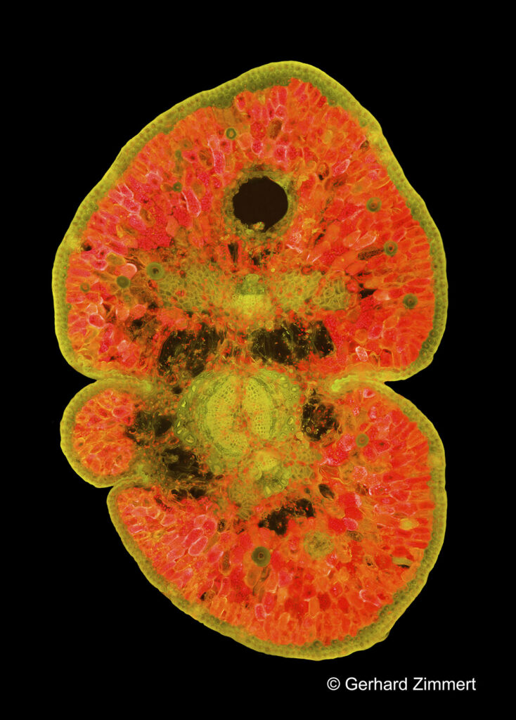
In the very last hours of the year, the first 13 images of the new series on needle cross-sections taken in autofluorescence*) have been completed. All the motifs are conifer cross-sections. The sections are between 35 and 50 μ thick. The images (panoramas from 2 to 7 stacks) can consist of up to 700 individual images. In order to be able to recognise the many details recorded, the images would have to be printed out at a size of more than 1 m, which is unfortunately not possible on the Internet. However, there will certainly be exhibition pictures from this series.
The exciting thing for me personally about this series is that I would not have been able to realise it in this quality a year ago. This is partly due to the new sensor technology (Nikon Z6) and a lens system in the microscope's UV beam path, which focusses the light onto a small diameter and thus concentrates it. These technical improvements enabled me to tackle two aspects of a problem (1) the long exposure time, which was originally in the area of 30 seconds for single images, and (2) the decrease in autofluorescence activity with prolonged excitation by UV light. If you only take single images, you don't necessarily notice the decrease in autofluorescence immediately, but with a panoramic image (series) the activity must be stable. With my equipment, I currently only need 1 to 2 seconds per individual image, which massively reduces the processing time and the first stack does not differ from the last (even if a stack consists of up to 100 individual images).
Technical data on the images: Objectives (UPLSAPO with 4x, 10x, 20x and 40x) were used - these are further amplified in the photo beam path by a 2.5x magnification of the projective lens and an additional effective tube magnification.
Illumination was with a 100 watt HBO lamp in the APO lamp house, the prism used for all images is blue (B) wideband.
*) Autofluorescence: the object is excited with light (in my case above blue) and emits light back towards the sensor in the visible range.